Amygdalostriatal transition zone identified in prolonged threat response
Neuroscientists have pinpointed a small brain region, the amygdalostriatal transition zone (ASt), that maintains neural activity for the full duration of a threat cue.
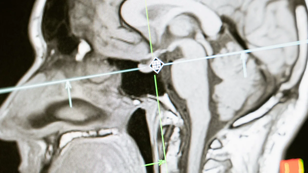
The amygdalostriatal transition zone (ASt), a small cluster of brain cells between the amygdala and striatum, has been identified as the region that maintains continuous neural activity for the full duration of a threat cue. This discovery fills a long-standing gap in understanding how brief threat signals translate into lasting defensive behaviors relevant to human anxiety disorders. Unlike amygdala neurons, which fire only briefly when a threat first appears, ASt neurons fire with both strength and longevity in response to danger cues, and direct stimulation of these cells drives freezing and avoidance behaviors in mice.
ASt neurons drive defensive behavior through specific cell types
Researchers used optogenetics to control specific neurons using light in 18 genetically engineered mice. When light was shined into the ASt, mice immediately began to freeze. In a separate test, mice actively avoided the side of an enclosure that triggered the laser. The team then tested 35 additional mice to isolate the effects of two primary cell types in the ASt.
| Neuron Type | Percentage in ASt | Effect When Activated |
|---|---|---|
| Drd2-positive | ~71% | Produces freezing and avoidance behaviors |
| Drd1a-positive | ~26% | No effect on defensive behavior |
Activating only Drd2-positive neurons produced freezing and avoidance, while activating Drd1a-positive neurons had no effect. Observations of calcium levels showed Drd2-positive neurons maintained high, sustained activity throughout threat-predicting tones. When the team silenced Drd2-positive ASt neurons in a group of 21 mice, defensive behaviors like freezing or sudden darting were reduced by nearly 50 percent. Blocking these cells did not change responses to reward cues or alter general movement.
Experimental evidence links ASt activity to learned threat cues
In experiments with 15 live mice, the animals were trained in an environment where a 20-second auditory tone predicted a mild foot shock, while a different 20-second tone predicted a sweet liquid reward. ASt neurons responded strongly to the shock-predicting tone and fired continuously for the entire 20-second duration. Trials with the highest defensive behavior, such as freezing, were accompanied by ASt firing rates over three times higher than in trials with low defensive behavior. The researchers tracked this using miniature microscopes attached to the mice's heads. Mice did not show ASt activation when placed in a context associated with fear, only when they heard the specific warning tone. The experiments used only a single type of threat and a single type of reward.
Implications for understanding and treating fear-related disorders
The ASt is described as a 'missing piece' of fear circuits that was hiding in plain sight for decades. Scientists hope to investigate whether this pathway plays a role in conditions like anxiety or post-traumatic stress disorder, where responses to external stimuli are disrupted. The findings may inform future therapeutic development for fear and panic disorders, including PTSD, panic disorder, and anxiety, which affect hundreds of millions of people globally. Fergil Mills, a researcher involved in the work, noted the feeling that led to the discovery: "It felt as though an important part of the neural circuitry had yet to be found." The ASt may serve as a critical circuit component in disorders where sustained fear responses to cues are maladaptive.