Duke's Injectable Scaffold Aids Stroke Recovery in Mice
Duke University researchers have developed an injectable biomaterial that, in mice, transformed stroke-damaged brain cavities into environments that
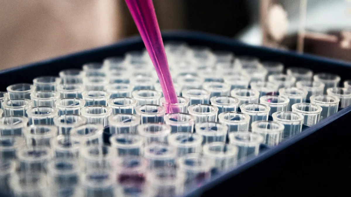
Biomedical engineers at Duke University have created an injectable treatment that helped mouse brains rebuild after a stroke. The therapy transformed the cavity left by dead tissue into a scaffold that recruited immune cells, spurred new blood vessel growth, and led to recovered movement.
According to the study published in Cell Biomaterials, the approach focuses on healing the damage that remains after emergency clot removal. "Once brain tissue has been lost, restoring blood flow is no longer enough," said Tatiana Segura, the Robert Plonsey Distinguished Professor of Biomedical Engineering at Duke. The team's goal was to engineer the injured space to kickstart a coordinated repair process.
Engineering a Healing Environment
The treatment uses a material called a microporous annealed particle scaffold, or MAPS. It is made of hydrogel microparticles that assemble into a porous structure inside the stroke cavity. This framework gives cells a place to enter and work. The researchers then attached signaling molecules and extracellular vesicles-tiny packages released by brain cells called astrocytes-to the scaffold's surface. This kept the repair signals concentrated where they were needed.
"We are not simply placing a material into the brain," Segura explained. "We are engineering a local environment that can coordinate several parts of the repair response." One combination of signals, IL-4 and C1q, proved especially good at drawing helpful immune cells into the damaged area.
A Surprising Role for Immune Cells
The therapy recruited a mix of immune cells, including macrophages and a persistent population of neutrophils. Neutrophils are typically linked to harmful inflammation early after a stroke. However, the Duke study suggests their role can change. In this engineered environment at a later stage, neutrophils appeared to support healing.
When researchers reduced the neutrophil population, blood vessel formation declined and the scaffold remodeled less. "This result changes how we think about neutrophils after stroke," said lead scientist Shangjing Xin. "Their role appears to depend on when they arrive, where they are located, and the signals they receive."
Measurable Improvements in Recovery
The biological changes led to functional gains. Mice treated with the optimized scaffold performed better on a grid-walking test that measures forelimb placement errors. By eight weeks, their performance was statistically indistinguishable from healthy mice, and the improvement lasted for the rest of the study.
The scaffold was essential. Extracellular vesicles administered without it failed to produce comparable blood vessel repair. The porous architecture and its ability to concentrate signals were critical.
A Preclinical Step Forward
The work remains in early stages. It has only been tested in mouse models, injected directly into the brain. Further research is needed on safety, mechanism, and scalability in larger models. The team is now investigating vesicles from human-derived astrocytes as a more clinically relevant source.
Segura likened the approach to ecological restoration. "You do not restore an ecosystem simply by containing the initial damage," she said. "You have to create the conditions that allow life to return."